Previous: 2.3. Knowledge domains for understanding.
3. Detecting signals in the eye and creating maps in the brain.
What can be seen with our eyes and is located in our brains?
This Chapter refers to the process of viewing that occurs when the tracks of bundles of light rays are concentrated in the human eye. We also consider the development of conceptual models in different environments by using positioning and orientation tools.
Learning objectives of Chapter 3.
After this Chapter you should be able to:
• Describe the Nobel Prizes in Medicine awarded in 1911, 1967 and 1981 concerning visual systems and processes.
• Describe the Nobel Lectures corresponding to the 2014 Nobel Prize in Medicine awarded for discovering a positioning system in the brain.
• Identify the cognitive procedures (Inquiring, Training, Comprehension, and Metacognition) in the description of models explaining the orbit of Mercury as presented by Kepler, Newton and Einstein.
Description of content of Chapter 3.
Section 3.1. Understanding the mechanisms of vision.
We comment on three Nobel Prizes in Physiology or Medicine: to Allvar Gullstrand in 1911 “for his work on the dioptrics of the eye” [Dioptrics is the study of the refraction of light, especially by lenses.], to Ragnar Granit, Haldan Keffer Hartline and George Wald in 1967 “for their discoveries concerning the primary physiological and chemical visual processes in the eye”, and to David H. Hubel and Torsten N. Wiesel in 1981 “for their discoveries concerning information processing in the visual system”.
Section 3.2. Description of a mental Global Positioning System (GPS).
We consider the 2014 Nobel Prize in Physiology or Medicine awarded to John O’Keefe, May-Britt Moser and Edvard I. Moser “for their discoveries of cells that constitute a positioning system in the brain”. Their investigations have served to understand the working conditions of a sort of mental GPS (Global Positioning System) used for visualization, memorization, and learning.
Section 3.3. Cognitive procedures for conceptual models.
We apply the Cognitive procedures for creating conceptual models (Inquiring, Training, Comprehension, and Metacognition) to the description of the motion of the planet Mercury around the Sun.
3.1. Understanding the mechanisms of vision.
Intelligence comes from Latin intus legere: to read inside. We need eyes to read, to see and understand shapes and meanings. Human eyes function like windows to observe the external world, communication channels for transmitting messages and interpretation screens to receive and process electromagnetic signals in a range of frequencies limited by ultraviolet and infrared radiations. We see because external stimuli are transformed into internal images. The sense of vision implies control of light in motion.
Eyes are the first elements in the chain eye-optic nerve-brain: the receiver, the transmitter, and the interpreter. This chain makes possible to distinguish, understand and employ representations of information and interpretations of knowledge about optical properties of objects. From the point of view of embryology eyes and brains come from the same type of cells.
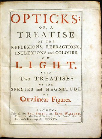
The study of Optics, as a branch of Physics, deals with three aspects of light: geometrical optics concerns propagation in straight lines, reflection, and refraction (see Figure I.8); physical optics refers to interference, diffraction and polarization, and quantum optics considers the behavior of photons and their interactions with different materials.

|

|
|---|
https://upload.wikimedia.org/wikipedia/commons/thumb/a/a0/Opticks.jpg/340px-Opticks.jpg
Figure I.8. Two communication productions concerning geometrical Optics developed in the XVIII Century: “Cover of the first edition of Newton's Opticks (1704)” (left image) and “Illustrations of various optical instruments from the 1728 Cyclopaedia (An Universal Dictionary of Arts and Sciences)” (right image).
In what follows we analyze three Nobel Prizes in Physiology or Medicine recognizing important contributions to the study of vision: the Prizes awarded in 1911 to Allvar Gullstrand, in 1967 to Ragnar Granit, Haldan Keffer Hartline and George Wald, and in 1981 to David H. Hubel and Torsten N. Wiesel. For each one of these six laureates we analyze two kinds of publications available in the web page of the Nobel Foundation: a full quotation of the document called WORK that explains the main contributions of each laureate and the description of their Nobel Lectures that we concentrate in a table containing two columns: the first one dedicated to Accepted knowledge or questions under discussion in laureate´s time and the second one regarding Laureate´s contributions or explanations.
1911 Medicine Nobel Prize awarded to Gullstrand.
WORK: "Our vision is based on the eye’s lens breaking up light from the outside world and converting it into an image at the back of the eye. From here, photosensitive retinal cells convert the light into nerve impulses that eventually become visual images. Calculating the path rays of light take through the eye and how an image is created is very complicated because the eye’s lens consists of different layers that refract light to different degrees. Moreover, the lens also changes shape. However, Allvar Gullstrand (1862-1930) succeeded in doing just that in the 1890s using advanced mathematics." (Figure I.9).

|

|
|---|
(Images credits: CC Wikimedia Commons: https://upload.wikimedia.org/wikipedia/commons/thumb/1/1e/Schematic_diagram_of_the_human_eye_en.svg/600px-Schematic_diagram_of_the_human_eye_en.svg.png https://api.openverse.org/v1/images/26b6ccee-dbe5-4408-8d26-7da1525c6b09/thumb/ and https://api.openverse.org/v1/images/d6ce0572-f1cc-4071-830a-05b20f58dbfe/thumb/)
Figure I.9. Structure of human eye (left image) and overview of a photosensitive retinal cell (right image). [ipRGCs means Intrinsically photosensitive retinal ganglion cells.]
MLA style: Allvar Gullstrand – Facts. NobelPrize.org. Nobel Prize Outreach AB 2023. Sat. 11 Mar 2023. https://www.nobelprize.org/prizes/medicine/1911/gullstrand/facts/
NOBEL LECTURE: How I Found the Mechanism of Intracapsular Accomodation by Gullstrand.
MLA style: Allvar Gullstrand – Nobel Lecture. NobelPrize.org. Nobel Prize Outreach AB 2023. Sat. 11 Mar 2023. https://www.nobelprize.org/prizes/medicine/1911/gullstrand/lecture/

1967 Medicine Nobel Prize awarded to Granit.
WORK: "Our vision works by the light around us being captured by a large number of light-sensitive cells located in the retinas at the back of our eyes. After a series of nerve switches and conversions of chemical and electrical signals, this results in visual impressions. Using very sophisticated electrodes, Ragnar Granit (1900-1991) was able to study the electrical impulses from the retina’s cells. In studies conducted from the 1930s to the 1950s, he demonstrated that there are different types of cones (the cells that enable color vision) and that these are sensitive to light of three different wavelengths." (Figure I.10).

|

|
|---|
(Images credits: CC Wikimedia Commons:
Figure I.10. Functional parts of two kinds of photosensitive cells in the retina (rods and cones) (left image) and anatomy of a cone cell (right image).
MLA style: Ragnar Granit – Facts. NobelPrize.org. Nobel Prize Outreach AB 2023. Sat. 11 Mar 2023. https://www.nobelprize.org/prizes/medicine/1967/granit/facts/
NOBEL LECTURE: The Development of Retinal Neurophysiology by Granit.
MLA style: Ragnar Granit – Nobel Lecture. NobelPrize.org. Nobel Prize Outreach AB 2023. Sat. 11 Mar 2023. https://www.nobelprize.org/prizes/medicine/1967/granit/lecture/

1967 Medicine Nobel Prize awarded to Hartline.
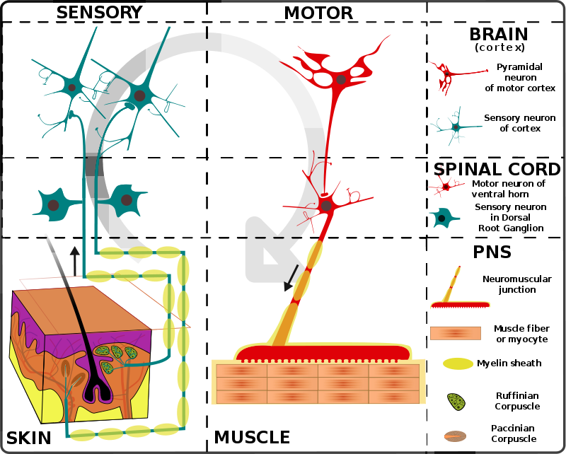
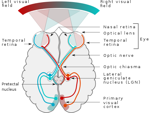
WORK: "Our vision functions because light from the surrounding world is captured by many light-sensitive cells in the retina at the back of the eye. A series of reconnections and transformations of chemical and electrical signals finally result in visual impressions. In studies of the horseshoe crab around 1950, Keffer Hartline (1903-1983) analyzed how the primary signals from visual cells are processed in a network of nerve cells. Among other things, he showed that when a cell is stimulated, signals from surrounding cells are suppressed. This makes it easier to understand the concept of contrasts." (Figure I.11).

|

|
|---|
(Images credits: CC Wikimedia Commons:
Figure I.11. Structure of the nerve system organization in humans (left image) and human vision pathway (right image). (PNS means peripheral nervous system.)
MLA style: Keffer Hartline – – Facts. NobelPrize.org. Nobel Prize Outreach AB 2023. Sat. 11 Mar 2023. https://www.nobelprize.org/prizes/medicine/1967/hartline/facts/
NOBEL LECTURE: Visual Receptors and Retinal Interaction by Hartline.
MLA style: Keffer Hartline – Nobel Lecture. NobelPrize.org. Nobel Prize Outreach AB 2023. Sat. 11 Mar 2023. https://www.nobelprize.org/prizes/medicine/1967/hartline/lecture/

1967 Medicine Nobel Prize awarded to Wald.
WORK: "Our vision functions because light from the surrounding world is captured by many light-sensitive cells in the retina at the back of the eye. George Wald (1906-1997) found that vitamin A is an important component in rhodopsin, a light-sensitive substance in the retina, and explained in a series of studies from the 1930s to the 1960s how light causes rhodopsin to change form and be converted. This conversion gives rise to signals in a complicated network of nerve cells by which a number of reconnections and transformations occur before the signals eventually are transformed into visual impressions in the brain."
MLA style: George Wald – Facts. NobelPrize.org. Nobel Prize Outreach AB 2023. Sat. 11 Mar 2023. https://www.nobelprize.org/prizes/medicine/1967/wald/facts/
NOBEL LECTURE: The Molecular Basis of Visual Excitation by Wald.
MLA style: George Wald – Nobel Lecture. NobelPrize.org. Nobel Prize Outreach AB 2023. Sat. 11 Mar 2023. https://www.nobelprize.org/prizes/medicine/1967/wald/lecture/

1981 Medicine Nobel Prize awarded to Hubel.
WORK: "Our vision works by the light around us being captured by a large number of light-sensitive cells located in the retinas at the back of our eyes. The light is converted into signals that are sent to the brain and there converted into visual impressions. David Hubel (1926-2013) and Torsten Wiesel clarified how this process works during the 1960s: In the cerebral cortex signals are analyzed in sequence by cells with the specific tasks of interpreting contrasts, patterns, and movements. They also showed that this ability develops in children during the initial period after birth."
MLA style: David H. Hubel – Facts. NobelPrize.org. Nobel Prize Outreach AB 2023. Sat. 11 Mar 2023. https://www.nobelprize.org/prizes/medicine/1981/hubel/facts/
NOBEL LECTURE: Evolution of ideas on the primary visual cortex, 1955-1978: a biased historical account by Hubel.
- Introduction
- Hierarchy of visual cells
- Hypercomplex cells
- Architecture
- Orientation columns
- Ocular dominance columns
- Relationship between columns, magnification and field size
MLA style: David H. Hubel – Nobel Lecture. NobelPrize.org. Nobel Prize Outreach AB 2023. Sat. 11 Mar 2023. https://www.nobelprize.org/prizes/medicine/1981/hubel/lecture/

1981 Medicine Nobel Prize awarded to Wiesel.
WORK: "Our vision works by the light around us being captured by a large number of light-sensitive cells located in the retinas at the back of our eyes. The light is converted into signals that are sent to the brain and there converted into visual impressions. Torsten Wiesel (1924) and David Hubel clarified how this process works during the 1960s: In the cerebral cortex signals are analyzed in sequence by cells with the specific tasks of interpreting contrasts, patterns, and movements. They also showed that this ability develops in children during the initial period after birth."
MLA style: Torsten N. Wiesel – Facts. NobelPrize.org. Nobel Prize Outreach AB 2023. Sat. 11 Mar 2023. https://www.nobelprize.org/prizes/medicine/1981/wiesel/facts/
NOBEL LECTURE: The postnatal development of the visual cortex and the influence of environment by Wiesel.
- Introduction
- Monocular deprivation
- The critical period
- Recovery from deprivation
- Normal development
MLA style: Torsten N. Wiesel – Nobel Lecture. NobelPrize.org. Nobel Prize Outreach AB 2023. Sat. 11 Mar 2023. https://www.nobelprize.org/prizes/medicine/1981/wiesel/lecture/

Next: 3.2. Generation of a mental Global Positioning System (GPS).